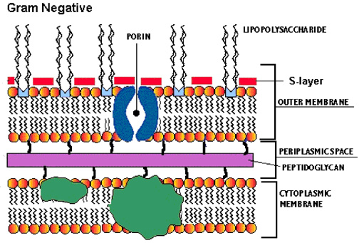
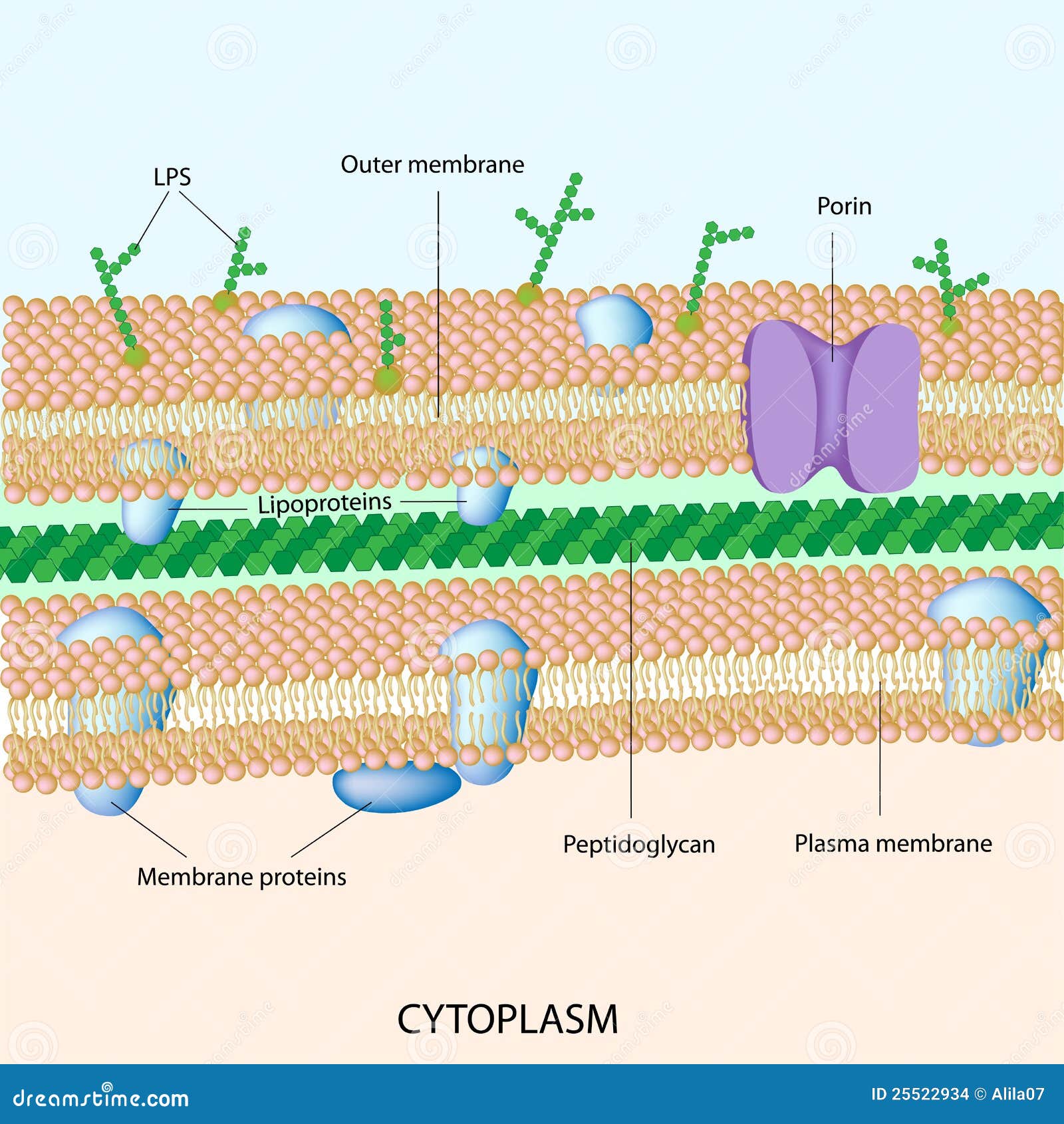
43 bacterial cell picture with labels
Interphase- Definition, Stages, Cell cycle, Diagram, Video The synthesis (S) phase is the phase of cell copying or cell duplication of its DNA of its entire genome. Gap 1 (G1) This is the phase in which the cell undergoes normal growth and cell function synthesizing high amounts of proteins. The cell increases in size and volume as more cell organelles are produced. What Is the Structure of a Bacterial Cell? (with pictures) The genetic information of a bacterial cell is encoded in a supercoiled structure of deoxyribonucleic acid ( DNA) suspended in the region known as the nucleoid. The bacterial chromosome is usually circular in shape. Other small pieces of DNA known as plasmids float independently in the cytoplasm apart from the main chromosome.
Parts of the Microscope with Labeling (also Free Printouts) 5. Knobs (fine and coarse) By adjusting the knob, you can adjust the focus of the microscope. The majority of the microscope models today have the knobs mounted on the same part of the device. Image 5: The circled parts of the microscope are the fine and coarse adjustment knobs. Picture Source: bp.blogspot.com.

Bacterial cell picture with labels
What is Streptococcus Bacteria? (with pictures) - The Health Board Streptococcus pneumoniae, or pneumococcus, is the most common cause of invasive bacterial infection of children and the elderly. It can cause pneumonia, sinusitis and meningitis, amongst other diseases. Streptococcus pneumoniae may cause either lobar pneumonia, which affects an entire lung lobe, usually in younger adults, or bronchial pneumonia ... Label the image to demonstrate your understanding of bacterial cell ... Label the image to demonstrate your understanding of bacterial cell structures. Advertisement ottahlisa814 is waiting for your help. Add your answer and earn points. Answer Expert Verified 0 ahsan57900 Thread like structures that are present at the center of bacterial cell is known as nucleoid which is a genetic material of that organism. What Does Vaginitis Look Like - Pictures of STDs - The STI Project Paavonen, Jorma, and Robert C. Brunham. "Bacterial vaginosis and desquamative inflammatory vaginitis." New England Journal of Medicine 379.23 (2018): 2246-2254. Vieira-Baptista, Pedro, et al. "Bacterial vaginosis, aerobic vaginitis, vaginal inflammation and major Pap smear abnormalities."
Bacterial cell picture with labels. Flagella: Structure, Arrangement, Function - Microbe Online Protozoa are a heterogeneous group that possesses three different organs of locomotion: flagella, cilia, and pseudopods. Certain protozoa, such as Leishmania and Trypanosoma have flagellated forms called promastigotes and non flagellated forms called amastigotes. Giardia lamblia and urogenital flagellate Trichomonas vaginalis also have flagella. Various shapes and arrangements of Bacterial cells Arrangements of Cocci. Arrangements of Bacilli (rod shaped bacteria) Spiral Bacteria. However pleomorphic bacteria can assume several shapes, following are the three basic bacterial shapes: Coccus (plural-cocci) : spherical. Bacillus (plural-bacilli) : rod-shaped. Spiral : twisted. Plasma Membrane (Cell Membrane) - Genome.gov Definition. The plasma membrane, also called the cell membrane, is the membrane found in all cells that separates the interior of the cell from the outside environment. In bacterial and plant cells, a cell wall is attached to the plasma membrane on its outside surface. The plasma membrane consists of a lipid bilayer that is semipermeable. What is a Recombinant Plasmid? (with pictures) - Info Bloom Several factors are involved in constructing a plasmid that can be used in molecular cloning. The plasmid must have a selectable marker. This makes it possible to select a cell with the gene. Normally, the population of cells lacking the gene with the marker greatly out-number the amount of cells that carry it.
Plant and Animal Cell: Labeled Diagram, Structure, Function - Embibe Plant Cell: Plant cells are eukaryotic cells with a true nucleus along with specialized structures called organelles that carry out certain specific functions. Animal Cell: An animal cell is a type of eukaryotic cell that lacks a cell wall and has a true, membrane-bound nucleus along with other cellular organelles. Learn the parts of a cell with diagrams and cell quizzes - Kenhub Prokaryotic cells: Simple, self-sustaining cells (bacteria and archaea) Eukaryotic cells: Complex, non self-sustaining cells (found in animals, plants, algae and fungi) In this article, we'll be focusing on eukaryotic cells. Two major regions can be found in a cell. The first is the cell nucleus, which houses DNA in the form of chromosomes. Microscopic Morphology - BIO 2410: Microbiology - Research Guides at ... The microscope images in this section show different bacterial structures visible using the light microscope. All images were photographed at 1000x magification. Structure 1 1. What bacterial structure appears as halos around the bacteria show here? 2. What staining procedure was used to prepare this specimen? Structure 1A Structure 2 Bacterial Cell Morphology and Classification: Definition, Shapes ... Shapes of Cells Bacteria The first shape is called coccus, plural cocci. Cells that have a cocci shape are spherical, resembling tiny balls. The 'strep' in strep throat actually refers to the...
Bacterial Capsule: Importance, Capsulated Bacteria - Microbe Online Capsule (also known as K antigen) is a major virulence factor of bacteria, e.g. all of the principal pathogens which cause pneumonia and meningitis, including Streptococcus pneumoniae, Haemophilus influenzae, Neisseria meningitidis, Klebsiella pneumoniae, Escherichia coli, and group B streptococci have polysaccharide capsules on their surface. Parts of a microscope with functions and labeled diagram Head - This is also known as the body. It carries the optical parts in the upper part of the microscope. Base - It acts as microscopes support. It also carries microscopic illuminators. Arms - This is the part connecting the base and to the head and the eyepiece tube to the base of the microscope. Bacterial Cytoplasm & Cell Membrane: Structure & Components A bacterial cell can increase surface area of the cell membrane with only a small change in the volume of the cytoplasm by making many invaginations of the cell membrane. 'Invagination' is just a ... Decoding a Probiotic Product Label - International Scientific ... Dietary supplement products formulated with untested microbes should similarly not be labeled as probiotics. Also, a label might not provide adequate information on what types of microbes are contained in the product. Genus and species may be listed, but no strain designation. Maybe only "bifidobacteria" or "lactobacilli" are listed.
Different Size, Shape and Arrangement of Bacterial Cells When viewed under light microscope, most bacteria appear in variations of three major shapes: the rod (bacillus), the sphere (coccus) and the spiral type (vibrio). In fact, structure of bacteria has two aspects, arrangement and shape. So far as the arrangement is concerned, it may Paired (diplo), Grape-like clusters (staphylo) or Chains (strepto).
Plant Cell Diagram - Diagrammatic representation of a generalized plant ... Animal cell picture with labels. A schematic diagram showing a simple layering process. Locate each organelle in the plant cell. This polysaccharide provides plant cells with strength and rigidity. Label the organelles in the diagram below. What structures are present in an animal cell, but not in a plant cell?
Bacteria Structure in Detail with Clear Diagram and Functions The bacterial cell structure has 70 s ribosomes. It consists of two sub-units as the 50S and 30S ribosomes. The ribosomes are present in the free-flowing form are grouped into polysomes. These ribosomes are useful in protein synthesis. But, humans, are the target of an antibiotic attack in the treatment of bacterial infections. Nuclear material
IT's All About A Bacterial Cell! - ProProfs Quiz 1. Bacteria can be which of the following shapes? A. Bacillus, Spirilia, Canadiaus B. Bacillus, Spirilia, Conpus C. Bacillus, Spirilia, Coccus D. Bacillus, Quadrain, Coccus E. Bacillus, Quadrain, Conus 2. A bacteria named Staphylobaccilus would be: (hint, what does staph mean and what does baccilus mean? A. Rod shapped and clustered B.
5 Steps of Gram Staining Procedure: How to Interpret the Results Now you are ready for the gram staining procedure. 5. Gram Staining. Place the heat-fixed smear on a staining tray. Flood your smear gently with crystal violet. Let it stand for one minute. Slightly tilt slide and rinse gently with tap water or distilled water using a wash bottle. Now flood the smear gently with Gram's iodine.
Blood Histology Slides with Description and Labeled Diagram The blood is a specialized connective tissue that is fluid and circulates through the vascular channel. In the blood histology slide, you will find different types of cells with their specific features. This might be a short article where I will show you all the cells from the blood microscope slide with a labeled diagram and actual pictures.
Bacterial Identification | 8 Methods & Tests In Microbiology This method is to determine the individual bacterial physical appearance. 1. Based on size Here bacteria are identified based on their physical size. Like small size or big one expressed in microns. For this bacteria are viewed under a microscope to view the size.
Incredible Images Reveal the Complex Face of Living Bacteria Like Never ... By University College London October 25, 2021 Microscopy image of a living E. coli bacterium, revealing the patchy nature of its protective outer membrane. A densely packed network of proteins is interrupted by smooth, protein-free islands (labeled by dashed lines in the inset). Credit: Benn et al. UCL
50 Striking Microscopic Images of Viruses and Bacteria 50 Striking Microscopic Images of Viruses and Bacteria March 07, 2022 1/50 Colorized transmission electron micrograph showing H1N1 influenza virus particles. Surface proteins on the virus particles...


Post a Comment for "43 bacterial cell picture with labels"